|
| Physics and Astronomy |
|
| Physics Home | Study here | Our Teaching | Our Research | Our Centres | News | Work here | EMPS |
|
Home › Research › Biomedical Physics › Multiphoton Imaging and Spectroscopy › Gallery › MacroPoly.html
Back to top
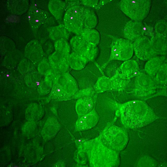
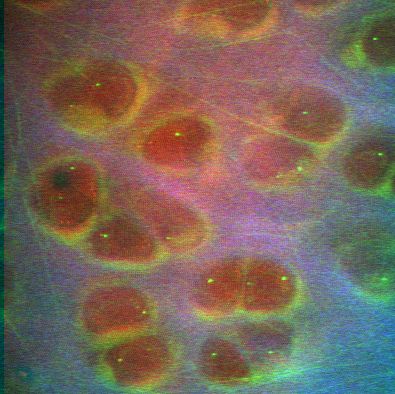
Macrophage uptake of 100nm polystyrene beadsImage by: Dr Julian Moger
CARS microscopy can be used to visualise polystyrene nanoparticles uptaken by live cells.
The figure shows 100 nm polystyrene beads in cultured macrophage cells; the vibrational signatures of the
nanoparticles and the cells can be separated by tuning (&omega p &omegas).
The green image contrast corresponds
to the CH2 of lipids (2485 cm-1) and the red C=C vibrational stretch of polystyrene (~1600 cm-1).
This study is facilitated by the strong Raman response and high density of
CH2 groups in lipids and C=C in polystyrene, which yield strong CARS signals.
|