| Physics and Astronomy |
|
| Physics Home | Study here | Our Teaching | Our Research | Our Centres | News | Work here | EMPS |
|
Biomedical Physics
Research Projects
Facilities
|
|
Back to top
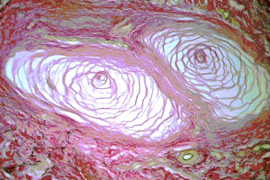
Measurements on Pacinian corpuscles in the fingertipIntroduction Micrograph of a Pacinian Corpuscle, (FOV 500×300 µm)
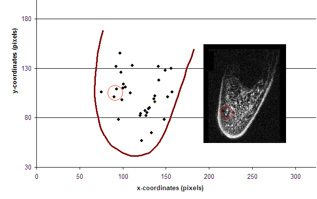
MRI investigation Identified locations within the right index distal
phalange. The U-shaped lines
indicate the outlines of the finger. The inset shows a single slices from the 3D
MRI data set.
Imaging was performed with a Philips whole-body imager at 1.5 T. A fat-suppression MRI technique was used to produce 3D data sets with a slice thickness of 140 µm and an in-plane resolution of 140 µm × 140 µm. The objects identified as Pacinian corpuscles are clustered off the midline of the fingertip, in close proximity to the expected locations of the digital nerves, matching the distributions described by Stark et al. [3]. Psychophysics investigation
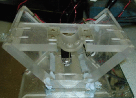
The vibrotactile stimulator, showing the curved surface on which the finger rests and the two stimulation sites within this surface; 
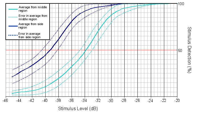
Psychometric curves - the full black line shows mean data from the side of the finger and the full grey line shows mean data from centre of the finger pad. The dotted lines indicate the ranges of standard error. Detection threshold (defined as 50% detection rate) was measured for 14 young-adult subjects. The mean threshold at the side of the finger was ~ 1.0 µm and at the midline was ~ 2.0 µm. The mean difference in threshold (defined as 50% detection rate) between the two sites was determined to be (5.4 ± 1.8) dB. ConclusionsThe MRI results indicate a non-uniform distribution of Pacinian receptors in the fingertip, with a lower density towards the centre of the fingerpad than toward the sides. The psychophysics results indicate a non-uniform vibrotactile sensitivity over the fingertip, with a higher threshold on the midline and a lower threshold toward the side. This lends support to the hypothesis that higher receptor density is associated with lower threshold, and vice versa.References
|
