My Research
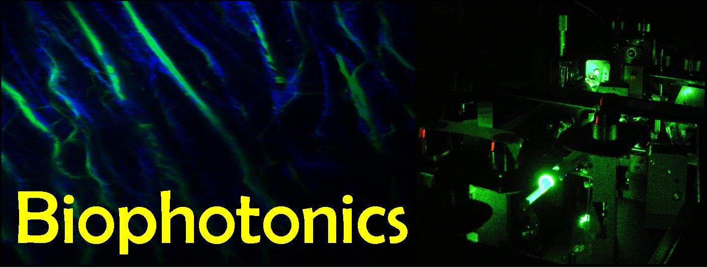
My research interests lie in the field of Biophotonics - the science of generating and harnessing light (photons) to image, detect and manipulate biological materials.
Currently I am focusing on the development and application of multiphoton techniques to aid the characterisation of biological tissues in both healthy and diseased states.
Multiphoton microscopy is a type of laser scanning microscopy that derives its contrast from `non- linear' optical properties of a sample. Non-linear imaging has many advantages over standard fluorescence confocal laser scanning microscopy,
with the principle advantages being an increase in depth penetration and stain-free molecular contrast.
Click here for a list of my current projects.
|
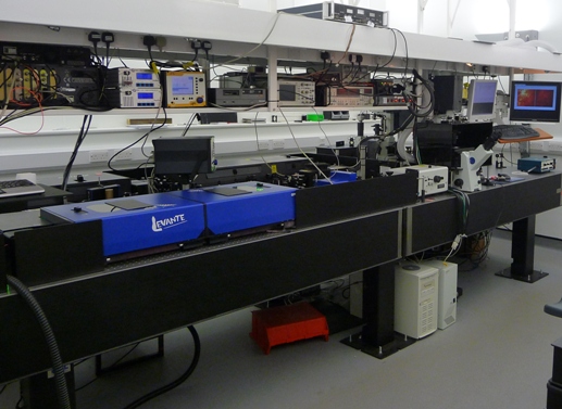
I have recently constructed a state-of-the-art multiphoton imaging spectroscopy laboratory, which is strongly supported by
staff with expertise in both the development and biological applications of multiphoton technology. The laboratory combines
multiple multiphoton contrast mechanisms in a single microscope, offering unrivalled flexibility allowing stain-free imaging
of multiple species within a sample. As well as providing non-invasive image contrast, multiphoton
interactions also contain spectroscopic information which can provide novel mechanisms for detection and characterisation of biomolecules in-vivo.
| 
|
|
|
|
|
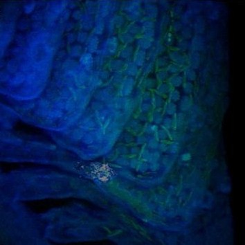
I am particularly interested in imaging the interaction of nanoparticles with biological systems. For example, I have used CARS microscopy to monitor the (eco)toxicological
impact of metal oxide nanoparticles in the aquatic environment. On the left, a 3D CARS image of a trout gill confirms that titanium oxide nanoparticles can become concentrated in the gill.
J. Moger, B. D. Johnston, C. Tyler. Imaging metal oxide nanoparticles in biological structures with CARS microscopy
Optics Express, 16 (5), 3408-3419, 2008. PDF
|
|
I work closely with the electromagnetic materials group to develop novel photonics techniques for bioanalysis using nanostructured surfaces.
|
|
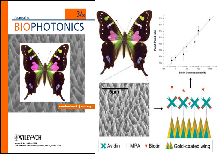
Naturally occurring nanostructures found on the wings of the Graphium butterfly can be used as substrates for surface-enhanced Raman scattering when coated with a thin film of gold or silver.
The substrates exhibit excellent biocompatibility and sensitivity, making them ideal for protein assaying. An assay using avidin/biotin binding showed that the substrates could be used to quantify protein binding directly from changes in the surface-enhanced Raman scattering (SERS)
spectra and were sensitive over a concentration range comparable with a typical enzyme-linked immunosorbent assays (ELISA) assay.
N.L. Garrett, P. Vukusic, F. Ogrin, E. Sirotkin, C. P. Winlove, J. Moger
Spectroscopy on the wing: Naturally Inspired SERS Substrates for Bio-Chemical Analysis
Journal of Biophotonics, Feb, 2009. PDF
| 
|
Back to top